
Discover effective approaches to integrative care for cardiorenal syndrome to better manage kidney and heart health.
Abstract
In this educational post, I will guide you through an evidence-based journey into the complex world of cardiorenal syndrome—the intricate crosstalk between the heart and kidneys that drives congestion, renal injury, and the progression of heart failure. We will explore the latest findings from leading researchers, moving beyond outdated models to a more nuanced understanding of how these two vital organs influence each other. I will break down complex physiological concepts like venous congestion, the critical roles of the right and left ventricles, and the paradigm shift from a simple “pre-renal” state to a more accurate “veno-renal” state.
This comprehensive guide covers the full spectrum of care, from initial diagnostic workups—including lab strategies (CMP, BNP, lactate), imaging, and physical assessment—to advanced management. I detail the pharmacology and practical use of diuretics, emphasizing thresholds, ceilings, and sequential nephron blockade. We will also discuss the role of inotropes, ultrafiltration, and mechanical circulatory support in refractory cases.
Throughout, I will explain how our multidisciplinary team at Injury Medical Clinic PA (Mission Plaza Injury Medical Clinic) in El Paso, Texas, operates. Under the medical direction of Dr. Maria Guadalupe Cardenas, MD, an internist with over 40 years of experience, we integrate integrative chiropractic care, functional medicine, and rehabilitation with guideline-directed medical therapy. This post will show how we align these modalities to improve hemodynamics, enhance decongestion, and restore function safely and effectively for patients navigating the cardiorenal landscape.
Our Integrative Approach at Injury Medical Clinic
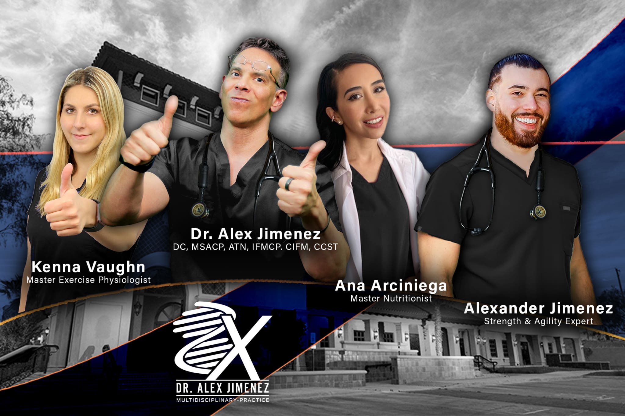
At Injury Medical Clinic PA, also known as Mission Plaza Injury Medical Clinic, in El Paso, Texas, we pride ourselves on a collaborative, multidisciplinary model of care. I am Dr. Alex Jimenez, and my extensive training in chiropractic (DC), nursing (APRN, FNP-BC), and functional medicine (CFMP, IFMCP) allows me to view patient health through a holistic lens. Our practice is further strengthened by the invaluable expertise of our Medical Director and Collaborative Physician, Dr. Maria Guadalupe Cardenas, MD. Dr. Cardenas is Board Certified in Internal Medicine (NPI #1164426749, Texas MD License #J2933) and brings over 40 years of clinical experience to our team.
This integrated setup, in which an MD provides medical direction alongside a chiropractor, is fundamental to our approach to complex cases, including those involving cardiorenal syndrome. It is a model common in many integrative and injury care clinics. Dr. Cardenas provides essential medical oversight, diagnosis, protocol guidance, and management of systemic diseases. At the same time, my role focuses on addressing the musculoskeletal, neurological, and biomechanical aspects through integrative chiropractic care, rehabilitation, and functional medicine principles.
This synergy allows us to address the root causes of dysfunction, not just the symptoms. For instance, while managing the medical aspects of heart failure, we can simultaneously use chiropractic adjustments and rehabilitation to improve thoracic mobility, enhance diaphragmatic function, and reduce the physical stress on a body already struggling with fluid overload and systemic inflammation. This comprehensive approach ensures all facets of a patient’s health are considered and treated cohesively, from personal injury care and rehabilitation to complex cardiometabolic strategies.
My Clinical Perspective: Why Cardiorenal Syndrome Matters
I practice integrative, evidence-based care with a focus on complex systems biology. In the clinic, patients seldom present with single-organ illnesses; they present with interconnected syndromes. Cardiorenal syndrome is one of the most striking examples of this reality. When the heart struggles to maintain adequate output, and the kidneys become congested or under-perfused, a self-reinforcing cycle of inflammation, fibrosis, hormonal imbalance, and fluid overload accelerates.
- Cardiorenal syndrome is the expression of a multidirectional crosstalk between the heart and kidneys.
- The renin-angiotensin-aldosterone system (RAAS) and natriuretic peptides act as opposing endocrine levers.
- Sympathetic nervous system overactivation, reactive oxygen species, and cytokine-driven fibrosis convert short-term compensation into long-term maladaptation.
- Effective care requires integrative thinking: we combine medical oversight, chiropractic mobilization, functional medicine nutrition, rehabilitation, and principles of injury care to reduce congestion and restore function safely.
Clinical leadership and oversight are essential. At Injury Medical Clinic PA, Dr. Maria Guadalupe Cardenas, MD, brings decades of internal medicine expertise to ensure safety and proper medical governance in a multidisciplinary environment. Under her direction, I integrate chiropractic and functional approaches that align with cardiometabolic goals, focusing on movement, vascular health, and autonomic recalibration.
The Endocrine Tug of War Between Heart and Kidneys
The heart and kidneys are potent endocrine organs engaged in a dynamic push-pull:
- The heart secretes atrial natriuretic peptide (ANP), B-type natriuretic peptide (BNP), and C-type natriuretic peptide (CNP). These signals favor vasodilation and natriuresis, promoting sodium and water excretion to reduce filling pressures and wall stress (Maisel et al., 2002).
- The kidneys initiate RAAS: renin converts angiotensinogen to angiotensin I, then angiotensin II drives vasoconstriction, and aldosterone promotes sodium and water retention—thereby raising preload and blood pressure (Hall, 2016; Zannad et al., 2012).
As heart failure evolves and renal perfusion fluctuates, RAAS predominates. BNP and NT-proBNP rise—often not merely as markers of stretch, but as endocrine signals attempting to counter RAAS. Think of this much like TSH in hypothyroidism: elevated because the system is trying, unsuccessfully, to restore balance. Over time, the kidneys’ endocrine power wins, and RAAS dominance amplifies congestion and fibrosis (Braunwald, 2015).
Why this matters clinically:
- Elevated NT-proBNP suggests not only volume or pressure overload but also systemic endocrine distress.
- RAAS predominance predicts worse outcomes, more renal injury, and diuretic resistance.
- Therapeutic strategies must aim to reduce RAAS activation, improve circulating volume, and, where possible, enhance natriuretic signaling.
A Paradigm Shift: From Forward Flow to Backward Congestion
For decades, the prevailing wisdom in heart failure management centered on contractility and forward flow. The thinking was that if we could make the heart pump stronger, everything else would fall into place. High “filling pressures”—the pressures inside the heart chambers before they contract—were seen as a necessary evil to maintain cardiac output. More recently, the spotlight has shifted to a long-overlooked player: the right ventricle (RV). We have historically under-recognized the profound impact of venous pressure. When the right side of the heart can’t effectively pump blood forward, pressure backs up throughout the entire venous system. This is the essence of venous congestion.
In my clinical observations, this manifests in several ways. We see fluid accumulate in the:
- Liver and Spleen: This leads to rapid enlargement of these organs, a condition known as splenomegaly.
- Abdominal Cavity: Fluid packs around the intestines and within the abdominal wall itself. On a CT scan, you can often see significant abdominal wall edema.
- Blood Vessels: The vessels become engorged. On echocardiography, we often observe that the inferior vena cava (IVC) is markedly “plump” and fails to collapse normally during inspiration.
This systemic fluid overload creates two major problems for a struggling heart:
- Increased Circulatory Volume: The total volume of blood the heart has to pump increases significantly.
- Increased Venous Congestion: The pressure within the venous system skyrockets. This back-pressure has profound implications for every organ, especially the kidneys.
The Kidney Under Pressure: Introducing the Veno-Renal State
Now, let’s connect this back to the kidneys. The kidneys are remarkable filters that operate on a pressure gradient. Think of the glomerulus, the kidney’s primary filtering unit, as a sophisticated sieve. High-pressure arterial blood flows in and exits into a low-pressure venous system. The difference between the high inflow pressure and the low outflow pressure creates the gradient that drives filtration. As venous pressure rises due to systemic congestion, the outflow pressure from the kidney increases. This narrows the pressure gradient.
When the gradient narrows, flow through the glomerulus slows down, and the kidney’s ability to filter waste is drastically reduced. We used to call this “pre-renal” failure, implying the problem was simply a lack of blood flow to the kidneys. Today, we are moving toward a more precise understanding: the veno-renal state. This concept emphasizes that it’s not just about forward flow; it’s equally, if not more, important to decongest the kidney by lowering venous pressure.
From Compensation to Maladaptation: SNS, Cytokines, and Fibrosis
Short-term compensation can be lifesaving, but the chronic state is different.
- Sympathetic nervous system (SNS) overactivation initially increases heart rate and contractility, but later drives myocardial remodeling, arrhythmias, and oxygen demand. It also propagates inflammatory cytokines and reactive oxygen species (ROS) that injure the myocardium and kidneys (Francis et al., 1990; Cohn et al., 1984).
- Within the nephron, persistent inflammatory signaling leads to glomerular and interstitial damage, sclerosis, and fibrosis. Distal tubular vacuolization impairs sodium and water handling, progressively reducing renal function (Ronco et al., 2010).
- Clinically, these changes produce diuretic resistance, worsening edema, and a trajectory of declining eGFR despite efforts to decongest (Damman et al., 2014).
Why this matters:
- Aggressive but nuanced decongestion is necessary to reverse venous hypertension and relieve renal congestion.
- We must use guideline-directed medical therapy (GDMT)—beta-blockers, ACE inhibitors/ARBs/ARNI, mineralocorticoid receptor antagonists (MRAs), and SGLT2 inhibitors—tailored to renal function, potassium, and blood pressure.
- Addressing intra-abdominal and splanchnic congestion is often the turning point in restoring effective diuresis.
Hemodynamics, Forward vs. Backward Flow, and Venous Reservoirs
In heart failure, reduced stroke volume and increased filling pressures lead to decreased cardiac output and elevated preload. While ankle edema is a common sign, it is often a late one. Earlier and more impactful, fluid accumulates within the splanchnic venous reservoir: the vascular bed of the liver, spleen, and omentum. Elevated right-sided pressures expand this reservoir, causing abdominal congestion that:
- Raises intra-abdominal pressure.
- Compresses renal veins and ureters.
- Impairs gut perfusion and barrier integrity, fueling systemic inflammation.
- Reduces diuretic absorption with oral agents, worsening diuretic resistance (Mullens et al., 2009; Verbrugge et al., 2013).
Why this matters:
- Physical examination should emphasize jugular venous distention, hepatic jugular reflux, abdominal distention/ascites, and hepatomegaly.
- Imaging can assess IVC diameter, hepatic congestion, and portal vein pulsatility as markers of venous hypertension.
- Therapeutic emphasis shifts toward reducing right-sided pressures and mobilizing splanchnic venous volume.
Assessing Kidney Function and Building Context
When a patient with heart failure presents with dyspnea and worsening kidney function, our first step is a thorough evaluation. It’s crucial not to react to a single lab value in isolation. The first question I always ask is: What is this patient’s baseline renal function? A creatinine of 1.9 mg/dL may sound alarming, but if their history shows a chronic baseline of 1.7-1.8 mg/dL, it’s not a sudden acute kidney injury (AKI); it’s likely an acute-on-chronic issue. Knowing the baseline is vital for setting realistic treatment goals.
While creatinine is a common marker, I increasingly rely on the Glomerular Filtration Rate (GFR), which gives a much better picture of overall kidney function. Most modern heart failure therapies can be safely started in patients with a GFR above 30 mL/min, and some, like SGLT2 inhibitors, as low as 20 mL/min.
Modern Lab Strategy for Diagnostic Clarity
I use a systematic lab approach to clarify the interplay between the heart, kidneys, and liver and to risk-stratify perfusion status.
- Comprehensive Metabolic Panel (CMP): I order a CMP because it includes liver indices. If renal function is elevated but liver indices (AST, ALT, bilirubin) are normal, I question if the issue is intrinsic renal pathology rather than pure hemodynamic congestion. Conversely, elevated hepatic enzymes can indicate hepatic congestion due to right-sided heart failure (Damman et al., 2010).
- Complete Blood Count (CBC): This identifies anemia (worsening dyspnea) and leukocytosis (suggesting infection). I’ve seen patients present with extreme shortness of breath whose underlying problem was severe anemia (hemoglobin of 5 g/dL), not just heart failure.
- BNP or NT-proBNP: I order these to quantify cardiac wall stress. Elevated natriuretic peptide levels help confirm the presence and severity of heart failure and are strong predictors of outcomes (Januzzi et al., 2018).
- Lactate: I always get a lactate. Elevated lactate is a marker of poor perfusion. It helps me risk-stratify: a congested patient with normal lactate may be managed with careful diuresis, while one with elevated lactate (malperfusing) requires consideration of inotropes or mechanical support.
- Troponin: Significant elevation suggests acute myocardial infarction; mild elevations may reflect demand ischemia. This helps triage coronary evaluation.
- Urinalysis with Microalbumin: Gross proteinuria points to nephrotic syndrome or glomerulonephritis, which can masquerade as heart failure. Microalbuminuria helps detect underlying chronic kidney disease (CKD).
Imaging and Cardiac Testing: When and Why
- Echocardiography: If there has been no echo in the last 6 months, I order a repeat. It reveals ejection fraction (EF), diastolic function, RV performance, and pulmonary pressures.
- Renal Ultrasound: In suspected AKI, I consider renal ultrasound to rule out post-obstructive causes like hydronephrosis. Overlooking obstruction can lead to irreversible injury.
- 12-Lead ECG: I obtain an ECG to evaluate for ischemia and arrhythmias, such as atrial fibrillation, which can precipitate decompensation.
Physical Assessment: Functional Status and Congestion
I anchor my evaluation in the New York Heart Association (NYHA) functional classification because it aligns symptoms with functional capacity (Class I-IV). For congestion signs, I use patient-centered questions:
- Orthopnea:”“How many pillows do you use?” or “Are you sleeping in a chair?”
- Paroxysmal Nocturnal Dyspnea (PND): “Do you wake suddenly at night feeling you can’t breathe or panicked?” This is often misdiagnosed as panic attacks.
- Bendopnea: I observe patients tying their shoes and note the recovery pause. It is a validated sign of elevated filling pressures.
- Dyspnea on Exertion: I ask about functional tasks: “Can you walk across a parking lot?” or “Can you push a vacuum?”
I also assess early satiety, abdominal bloating, and peripheral edema. For malperfusion, I watch for fatigue, intermittent confusion, and oliguria.
Beating the Odds: “Conquering Congestive Heart Failure”- Video
Hemodynamic Profiles and Cardiorenal Phenotypes
Understanding the hemodynamic phenotype directs therapy:
- Warm and wet: Congested but perfusing. Focus on diuresis and vasodilators.
- Cold and wet: Congested with poor perfusion. Consider inotropes and mechanical support.
- Warm and dry: Perfusing without congestion. Optimize GDMT.
- Cold and dry: Poor perfusion without congestion. Cautious fluid challenges or inotropes.
We also recognize five cardiorenal phenotypes (Types I-V), which help determine whether to decongest the heart, protect the kidneys, or treat a systemic driver.
Loop Diuretics: Precision Dosing and Overcoming Resistance
Loop diuretics are central to decongestion. I largely favor torsemide or bumetanide over oral furosemide due to their predictable bioavailability.
- Relative potency (oral): 40 mg furosemide ≈ 20 mg torsemide ≈ 1 mg bumetanide.
- Oral-to-IV conversions: Furosemide is roughly 2:1 (80 mg oral ≈ 40 mg IV), while torsemide and bumetanide are 1:1.
- Dosing strategy: Start with an effective dose—often equal to or greater than the total daily home dose given IV. Schedule doses to align with waking hours to reduce nocturia and fall risks.
- Thresholds and Ceilings: The threshold (minimum effective dose) rises with renal impairment and edema. The ceiling is the point where more drug only adds side effects. When reaching the ceiling, I add a second agent.
- Pushes vs. Drips: The DOSE trial showed similar outcomes for bolus (push) versus continuous infusion strategies when dosing was correct (Felker et al., 2011). I use bolus dosing for most patients and continuous infusion for severe diuretic resistance.
A modest rise in creatinine (up to 0.5 mg/dL) after diuretic use often reflects RAAS activation rather than true AKI. Contextualizing this change prevents premature cessation of diuretics.
Sequential Nephron Blockade and Advanced Therapies
When loop diuretics are insufficient, I implement sequential nephron blockade by adding a thiazide (e.g., metolazone) 30-60 minutes before the loop diuretic to block distal sodium reabsorption. This requires vigilant electrolyte monitoring. Other key agents include:
- SGLT2 inhibitors (dapagliflozin, empagliflozin): These promote osmotic diuresis and confer renal and heart failure benefits even at lower eGFR thresholds (McMurray et al., 2019; Packer et al., 2020).
- Mineralocorticoid receptor antagonists (MRAs) (spironolactone, eplerenone): These counter aldosterone’s effects on sodium retention and fibrosis.
Applying Guideline-Directed Medical Therapy Safely
As we decongest, I prioritize GDMT with vigilant renal oversight:
- ACE inhibitors/ARBs/ARNI: Initiate with careful monitoring of creatinine and potassium. A small bump in creatinine is often acceptable if congestion is improving.
- Beta-blockers: Titrate slowly after volume status stabilizes.
- MRAs: Monitor potassium closely, especially in CKD.
- SGLT2 inhibitors: Can be started at eGFR ≥ 20 and improve outcomes across the board (Heerspink et al., 2020).
These steps, coordinated by Dr. Cardenas, align with contemporary guidelines (Heidenreich et al., 2022; McDonagh et al., 2021).
Inotrope Support and Mechanical Intervention
For patients with refractory oliguria despite optimal diuretics, inotropes can improve cardiac output and renal perfusion.
- Milrinone: A phosphodiesterase-3 inhibitor that provides inotropy and potent vasodilation, reducing RV afterload and improving the renal vein gradient. It is renally cleared, so I start at low doses.
- Dobutamine: A beta-adrenergic agonist that increases contractility, useful for augmenting contractile strength.
For severe fluid overload and diuretic resistance, ultrafiltration or dialysis can unload the venous system without further stimulation of the RAAS. In select cases of severe ventricular dysfunction, mechanical circulatory support (e.g., Impella, Protek Duo, ECMO) can bridge to recovery.
Integrative Chiropractic Care in Cardiorenal Management
In our practice, integrative chiropractic care is woven into a comprehensive clinical framework under medical oversight. Here is how it fits safely and effectively:
- Thoracic Cage Mobility and Rib Mechanics: Improving rib motion enhances respiratory mechanics, reduces the work of breathing, and supports venous return. Better diaphragmatic excursion can facilitate lymphatic and venous drainage.
- Diaphragmatic and Lymphatic Pump Techniques: Gentle, evidence-informed mobilizations support lymphatic flow, which can reduce interstitial fluid and augment decongestive strategies.
- Autonomic Balancing: With targeted manual therapy and breath retraining, we aim to reduce sympathetic overdrive and improve vagal tone, attenuating the endocrine-inflammatory cascade (Thayer & Lane, 2000). This complements beta-blocker therapy.
- Postural and Fascial Strategies: Addressing thoracolumbar fascial tension optimizes venous return from the lower body, reducing pooling and promoting splanchnic decongestion.
- Movement Prescriptions: Graded mobility and low-intensity exercise improve skeletal muscle pump function and microvascular flow, supporting diuretic efficacy.
- Safety Framework: All manual interventions are tailored to hemodynamic stability, anticoagulation status, and volume status. In significant decompensation, we focus on gentle mobilization, breathwork, and positioning.
Congestion is not merely a pharmaceutical problem. Movement, mechanics, and autonomic tone are powerful modulators of venous return and endocrine signaling.
Clinical Observations: Insights from Practice
From years of clinical care, including observations shared through my platforms at PushAsRx and LinkedIn, several patterns stand out:
- Abdominal congestion is the pivot point: When we treat the abdomen as a central venous reservoir—mobilizing the diaphragm and easing thoracic restrictions—patients often transition from diuretic resistance to responsiveness (Mullens et al., 2009).
- Subcutaneous furosemide can bridge gaps: In select patients, subcutaneous formulations offer predictable decongestion when IV routes are unavailable. Medical oversight is critical.
- Autonomic recalibration enhances GDMT tolerability: Breathing protocols and gentle manual therapy reduce sympathetic burden, improving tolerance to beta-blockers and RAAS inhibitors (Thayer & Lane, 2000).
- Functional strength matters: Restoring calf-pump efficacy and thoracic mobility reduces venous stasis, reduces edema, and improves quality of life.
Putting It All Together: A Practical Care Pathway
We organize care into coordinated steps:
- Assessment: Focused physical exam, POCUS, labs (NT-proBNP, electrolytes, renal function).
- Immediate Decongestion: IV loop diuretics, sequential nephron blockade if needed, strict I/O and electrolyte replacement.
- Guideline Therapy: Initiate and titrate GDMT under Dr. Cardenas’s oversight.
- Mechanic-Autonomic Integration: Chiropractic mobilizations, lymphatic techniques, graded movement, and breath training.
- Nutrition and Lifestyle: Individualized sodium/fluid strategy, protein optimization, and anti-inflammatory nutrition.
- Monitoring and Follow-up: Remote tracking, diuretic/GDMT adjustments, and coordination with specialists.
Cardiorenal syndrome demands systemic solutions. With Dr. Cardenas’s medical direction and our integrative team’s hands-on, evidence-based strategies, we help patients move from congestion and fatigue toward function and resilience.
References
- Bart, B. A., Boyle, A., Bank, A. J., et al. (2005). Ultrafiltration vs intravenous diuretics for patients hospitalized for acute decompensated heart failure. The Lancet, 366(9500), 135–141.
- Brater, D. C. (1998). Clinical pharmacology of loop diuretics. Hypertension, 31(6), 1130–1135.
- Braunwald, E. (2015). The path to an angiotensin receptor blocker–neprilysin inhibitor in the treatment of heart failure. New England Journal of Medicine, 373(11), 1099–1102.
- Cohn, J. N., Levine, T. B., Olivari, M. T., et al. (1984). Plasma norepinephrine as a guide to prognosis in patients with chronic congestive heart failure. New England Journal of Medicine, 311(13), 819–823.
- Damman, K., Navis, G., Voors, A. A., et al. (2010). Worsening renal function and prognosis in heart failure: Systemic congestion vs. low cardiac output. Journal of the American College of Cardiology, 55(12), 1218–1227.
- Damman, K., Valente, M. A., Voors, A. A., et al. (2014). Renal impairment, worsening renal function, and outcome in patients with heart failure. European Heart Journal, 35(7), 455–469.
- Felker, G. M., Lee, K. L., Bull, D. A., et al. (2011). Diuretic strategies in patients with acute decompensated heart failure. New England Journal of Medicine, 364(9), 797–805.
- Francis, G. S., Benedict, C., Johnstone, D. E., et al. (1990). Comparison of neuroendocrine activation in patients with left ventricular dysfunction with and without congestive heart failure. Circulation, 82(6), I–103.
- Hall, J. E. (2016). Guyton and Hall Textbook of Medical Physiology (13th ed.). Elsevier.
- Heidenreich, P. A., Bozkurt, B., Aguilar, D., et al. (2022). 2022 AHA/ACC/HFSA guideline for the management of heart failure. Circulation, 145(18), e895–e1032.
- Heerspink, H. J. L., Stefansson, B. V., Correa-Rotter, R., et al. (2020). Dapagliflozin in patients with chronic kidney disease. New England Journal of Medicine, 383(15), 1436–1446.
- Januzzi, J. L., Chen-Tournoux, A., & Christenson, R. H. (2018). Natriuretic peptide testing for heart failure. Clinical Biochemistry, 52, 1–15.
- Maisel, A., Krishnaswamy, P., Nowak, R. M., et al. (2002). Rapid measurement of B-type natriuretic peptide in the emergency diagnosis of heart failure. New England Journal of Medicine, 347(3), 161–167.
- McDonagh, T. A., Metra, M., Adamo, M., et al. (2021). 2021 ESC Guidelines for the diagnosis and treatment of acute and chronic heart failure. European Heart Journal, 42(36), 3599–3726.
- McMurray, J. J. V., Solomon, S. D., Inzucchi, S. E., et al. (2019). Dapagliflozin in patients with heart failure and reduced ejection fraction. New England Journal of Medicine, 381(21), 1995–2008.
- Miao, J., et al. (2020). Gut microbiota, hypertension and atherosclerosis. Frontiers in Physiology, 11, 142.
- Mullens, W., Abrahams, Z., Francis, G. S., et al. (2009). Importance of venous congestion for worsening of renal function in advanced decompensated heart failure. Journal of the American College of Cardiology, 53(7), 589–596.
- Mullens, W., Verbrugge, F. H., Martens, P., et al. (2022). Acetazolamide in acute decompensated heart failure with volume overload. New England Journal of Medicine, 387(13), 1185–1195.
- Packer, M., Anker, S. D., Butler, J., et al. (2020). Cardiovascular and renal outcomes with empagliflozin in heart failure. New England Journal of Medicine, 383(15), 1413–1424.
- Packer, M., Carver, J. R., Rodman, D., et al. (1991). Effect of milrinone on mortality in severe chronic heart failure. The New England Journal of Medicine, 325(21), 1468-1475.
- Rangrass, G., & Pinsky, M. R. (2020). The right ventricle: the Rodney Dangerfield of the heart. Critical Care, 24(1), 1-3.
- Ronco, C., McCullough, P. A., Anker, S. D., et al. (2010). Cardio-renal syndromes: report from the consensus conference of the Acute Dialysis Quality Initiative. European Heart Journal, 31(6), 703-711.
- Shankar-Hari, M., Phillips, G. S., Levy, M. L., et al. (2016). Developing a new definition and assessing new clinical criteria for septic shock. JAMA, 315(8), 775–787.
- Stevens, P. E., & Levin, A. (2013). Evaluation and management of chronic kidney disease: synopsis of the Kidney Disease: Improving Global Outcomes 2012 Clinical Practice Guideline. Annals of Internal Medicine, 158(11), 825-830.
- Thayer, J. F., & Lane, R. D. (2000). A model of neurovisceral integration in emotion regulation and dysregulation. NeuroReport, 11(3), 1–5.
- Thayer, J. F., Åhs, F., Fredrikson, M., Sollers, J. J., & Wager, T. D. (2010). A meta-analysis of heart rate variability and neuroimaging studies: Implications for HRV as an index of stress and health. Neuroscience & Biobehavioral Reviews, 33(2), 81–88.
- Tiu, P. R., DeMaria, A. N., & Chang, T. I. (2023). Subcutaneous furosemide for decompensated heart failure: A practical update. JACC: Heart Failure, 11(4), 345–356.
- Verbrugge, F. H., Dupont, M., & Mullens, W. (2013). Abdominal contributions to congestion in heart failure. European Heart Journal, 34(7), 446–454.
- Yancy, C. W., Jessup, M., Bozkurt, B., et al. (2017). 2017 ACC/AHA/HFSA Focused Update of the 2013 ACCF/AHA Guideline for the Management of Heart Failure. Circulation, 136(6), e137-e161.
- Zannad, F., McMurray, J. J. V., Krum, H., et al. (2012). Eplerenone in patients with systolic heart failure and mild symptoms. New England Journal of Medicine, 364(1), 11–21.
SEO tags: cardiorenal syndrome, heart failure, renal congestion, RAAS, natriuretic peptides, diuretic resistance, SGLT2 inhibitors, integrative chiropractic care, autonomic nervous system, splanchnic venous reservoir, abdominal congestion, multidisciplinary clinic, internal medicine oversight, El Paso Texas, Injury Medical Clinic PA, Mission Plaza Injury Medical Clinic, functional medicine, rehabilitation, personal injury care, NT-proBNP, diuretic strategies, kidney disease, venous congestion, Dr. Alex Jimenez, right ventricle, GFR, creatinine, acute kidney injury, chronic kidney disease, decompensated heart failure, veno-renal state, hemodynamics, dyspnea, NYHA classification, lactate, echocardiogram, renal ultrasound, milrinone, dobutamine, ultrafiltration, CRRT, hemodialysis, RV failure
Post Disclaimer *
Professional Scope of Practice *
The information herein on "Integrative Care Solutions for Cardiorenal Syndrome" is not intended to replace a one-on-one relationship with a qualified health care professional or licensed physician and is not medical advice. We encourage you to make healthcare decisions based on your research and partnership with a qualified healthcare professional.
Blog Information & Scope Discussions
Welcome to El Paso's Premier Fitness, Injury Care Clinic & Wellness Blog, where Dr. Alex Jimenez, DC, FNP-C, a Multi-State board-certified Family Practice Nurse Practitioner (FNP-BC) and Chiropractor (DC), presents insights on how our multidisciplinary team is dedicated to holistic healing and personalized care. Our practice aligns with evidence-based treatment protocols inspired by integrative medicine principles, similar to those found on this site and our family practice-based chiromed.com site, focusing on restoring health naturally for patients of all ages.
Our areas of multidisciplinary practice include Wellness & Nutrition, Chronic Pain, Personal Injury, Auto Accident Care, Work Injuries, Back Injury, Low Back Pain, Neck Pain, Migraine Headaches, Sports Injuries, Severe Sciatica, Scoliosis, Complex Herniated Discs, Fibromyalgia, Chronic Pain, Complex Injuries, Stress Management, Functional Medicine Treatments, and in-scope care protocols.
Our information scope is multidisciplinary, focusing on musculoskeletal and physical medicine, wellness, contributing etiological viscerosomatic disturbances within clinical presentations, associated somato-visceral reflex clinical dynamics, subluxation complexes, sensitive health issues, and functional medicine articles, topics, and discussions.
We provide and present clinical collaboration with specialists from various disciplines. Each specialist is governed by their professional scope of practice and their jurisdiction of licensure. We use functional health & wellness protocols to treat and support care for musculoskeletal injuries or disorders.
Our videos, posts, topics, and insights address clinical matters and issues that are directly or indirectly related to our clinical scope of practice.
Our office has made a reasonable effort to provide supportive citations and has identified relevant research studies that support our posts. We provide copies of supporting research studies upon request to regulatory boards and the public.
We understand that we cover matters that require an additional explanation of how they may assist in a particular care plan or treatment protocol; therefore, to discuss the subject matter above further, please feel free to ask Dr. Alex Jimenez, DC, APRN, FNP-BC, or contact us at 915-850-0900.
We are here to help you and your family.
Blessings
Dr. Alex Jimenez DC, MSACP, APRN, FNP-BC*, CCST, IFMCP, CFMP, ATN
email: [email protected]
Multidisciplinary Licensing & Board Certifications:
Licensed as a Doctor of Chiropractic (DC) in Texas & New Mexico*
Texas DC License #: TX5807, Verified: TX5807
New Mexico DC License #: NM-DC2182, Verified: NM-DC2182
Multi-State Advanced Practice Registered Nurse (APRN*) in Texas & Multi-States
Multistate Compact APRN License by Endorsement (42 States)
Texas APRN License #: 1191402, Verified: 1191402 *
Florida APRN License #: 11043890, Verified: APRN11043890 *
Verify Link: Nursys License Verifier
* Prescriptive Authority Authorized
ANCC FNP-BC: Board Certified Nurse Practitioner*
Compact Status: Multi-State License: Authorized to Practice in 40 States*
Graduate with Honors: ICHS: MSN-FNP (Family Nurse Practitioner Program)
Degree Granted. Master's in Family Practice MSN Diploma (Cum Laude)
Dr. Alex Jimenez, DC, APRN, FNP-BC*, CFMP, IFMCP, ATN, CCST
(Board Certified: Family Practice Nurse Practitioner—Multistate)*
(Licensed Nurse Practitioner & Chiropractor - Multistate)*
Clinical Director
Digital Business Card
Dr. Maria Cardenas, MD
(Board Certified: Internal Medicine)
(Licensed Medical Doctor)
Medical Director, Clinical Director & Collaborative Physician
NPI # 1164426749
MD License #: J2933
Licenses and Board Certifications:
MD: Medical Doctor
DC: Doctor of Chiropractic
APRNP: Advanced Practice Registered Nurse
FNP-BC: Family Practice Specialization (Multi-State Board Certified)
RN: Registered Nurse (Multi-State Compact License)
CFMP: Certified Functional Medicine Provider
MSN-FNP: Master of Science in Family Practice Medicine
MSACP: Master of Science in Advanced Clinical Practice
IFMCP: Institute of Functional Medicine
CCST: Certified Chiropractic Spinal Trauma
ATN: Advanced Translational Neutrogenomics
Memberships & Associations:
TCA: Texas Chiropractic Association: Member ID: 104311
AANP: American Association of Nurse Practitioners: Member ID: 2198960
ANA: American Nurse Association: Member ID: 06458222 (District TX01)
TNA: Texas Nurse Association: Member ID: 06458222
NPI: 1205907805
| Primary Taxonomy | Selected Taxonomy | State | License Number |
|---|---|---|---|
| No | 111N00000X - Chiropractor | NM | DC2182 |
| Yes | 111N00000X - Chiropractor | TX | DC5807 |
| Yes | 363LF0000X - Nurse Practitioner - Family | TX | 1191402 |
| Yes | 363LF0000X - Nurse Practitioner - Family | FL | 11043890 |
| Yes | 363LF0000X - Nurse Practitioner - Family | CO | C-APN.0105610-C-NP |
| Yes | 363LF0000X - Nurse Practitioner - Family | NY | N25929 |
Dr. Alex Jimenez, DC, APRN, FNP-BC*, CFMP, IFMCP, ATN, CCST
(Board Certified: Family Practice Nurse Practitioner—Multistate)*
(Licensed Nurse Practitioner & Chiropractor - Multistate)*
Clinical Director
Digital Business Card
Dr. Maria Cardenas, MD
(Board Certified: Internal Medicine)*
(Licensed Medical Doctor)*
Medical Director, Clinical Director & Collaborative Physician
NPI # 1164426749
MD License #: J2933