
Ultrasound PRP for Hip Impingement Recovery
Abstract
In this educational post, I walk you through an integrative, evidence-based approach to treating hip impingement and instability in a hypermobile dancer who presents with end-range pain and mechanical clicking. I explain how I evaluate the hip with ultrasound, differentiate labral pathology from instability-driven irritation, and perform an ultrasound-guided intra-articular injection using high-concentration platelet-rich plasma (PRP) combined with a plasma protein concentrate. I also describe how and why I limit injectate volume in the hip, the technical aspects of safe needle placement, and what constitutes an optimal in-joint flow profile. Most importantly, I contextualize this interventional step within a comprehensive, integrative chiropractic care plan that addresses kinetic-chain dysfunctions, neuromuscular control, fascial continuity, and recovery behaviors. Throughout, I reference current research on PRP biologics, hip instability biomechanics, and ultrasound guidance, and I connect these findings to my clinical observations from practice.
— Dr. Alexander Jimenez, DC, APRN, FNP-BC, CFMP, IFMCP, ATN, CCST

Understanding Hip Impingement and Hypermobility in Dancers: What I Look For
When a young dancer presents with hip pain, I immediately consider the unique interplay of range-of-motion demands, tissue laxity, and load exposure. In this case, the patient has a history of being hypermobile and reports pain at end ranges and mechanical clicking. This points me toward two related but distinct considerations:
- Femoroacetabular impingement (FAI) mechanics at extremes of motion
- Capsuloligamentous laxity and microinstability without a large labral tear
On ultrasound, I identify the femoral head centrally with the acetabulum superior-lateral. The triangular, hyperechoic structure at the acetabular rim is the labrum. In this case, no large labral tear is evident, though the image suggests irritation with microinstability. Microinstability means the hip lacks robust constraint at the capsule and supporting soft tissues, especially under repetitive end-range loads typical in dance.
- Why this matters: Hypermobility can allow the femoral head to translate excessively in the socket at end ranges, leading to repetitive labral and capsular irritation without a frank tear. The result is nociceptive input, synovitis, and reactive guarding that degrade performance and encourage maladaptive movement patterns.
- Physiological underpinnings: The hip capsule and iliofemoral ligament complex provide passive restraint. When these structures are lax, the neuromuscular system must upregulate dynamic stabilizers (gluteus medius/minimus, deep rotators) to maintain centralization of the femoral head. If neuromuscular control lags, the labrum and cartilage experience increased shear and compression. Over time, synovial inflammation, labral fraying, and vulnerability of the chondrolabral junction can manifest (Nepple et al., 2015; Kalisvaart & Safran, 2015).
- Clinical cues: Pain at end range, clicking, feelings of giving way, and position-dependent discomfort are hallmark signs. In dancers, provocative positions include deep flexion, internal rotation, abduction-adduction transitions, and forced turnout.
My clinical observations across sports and performing arts settings show that dancers often have excellent mobility but insufficient load sharing across the posterior chain, particularly when fatigue sets in. At these points, anterior capsular strain and labral compression increase. I reinforce control strategies to build endurance and precision at range limits.
Why I Chose High-Concentration PRP With Plasma Protein Concentrate
Biologic therapies like PRP leverage the body’s own growth factors and signaling molecules to modulate inflammation and promote tissue repair. In this case, I use a high-concentration PRP combined with a plasma protein concentrate:
- High-concentration PRP delivers elevated levels of platelet-derived growth factor (PDGF), transforming growth factor-beta (TGF-β), vascular endothelial growth factor (VEGF), insulin-like growth factor-1 (IGF-1), and epidermal growth factor (EGF). These mediators can support tenocyte and chondrocyte metabolism, synovial homeostasis, and local angiogenesis appropriate for repair (Fitzpatrick et al., 2017; Chahla et al., 2021).
- Plasma protein concentrate enriches fibrinogen and other plasma proteins that can serve as a scaffold for growth factor retention and gradual release. This may improve residence time and reduce efflux from the joint, which is especially valuable in joints with capsular laxity, where distribution can be broad (Everts et al., 2020).
- Why not large volumes? The hip joint is less tolerant of high volumes than the knee, which can accommodate larger volumes during viscosupplementation or lavage. Intra-articular pressure in the hip rises quickly, potentially provoking pain and pushing fluid into the periarticular planes. Concentrating the PRP allows therapeutic dosing in a small volume.
- My protocol in this scenario: 4 cc of high-concentration PRP plus 2 cc of plasma protein concentrate. The goal is to maximize anabolic signaling while minimizing capsular distension.
Physiologically, this combination aims to:
- Downregulate catabolic cytokines in synovium (e.g., IL-1β, TNF-α)
- Encourage matrix synthesis at the chondrolabral interface
- Enhance capsular tissue quality by supporting fibroblast activity
- Improve pain through modulation of peripheral nociceptor sensitization
This approach aligns with emerging evidence supporting PRP for hip intra-articular conditions and labral-related pathology when carefully selected and accurately placed (Dwyer et al., 2021; LaFrance et al., 2021).
Ultrasound-Guided Hip Injection: How I Ensure Accuracy and Safety
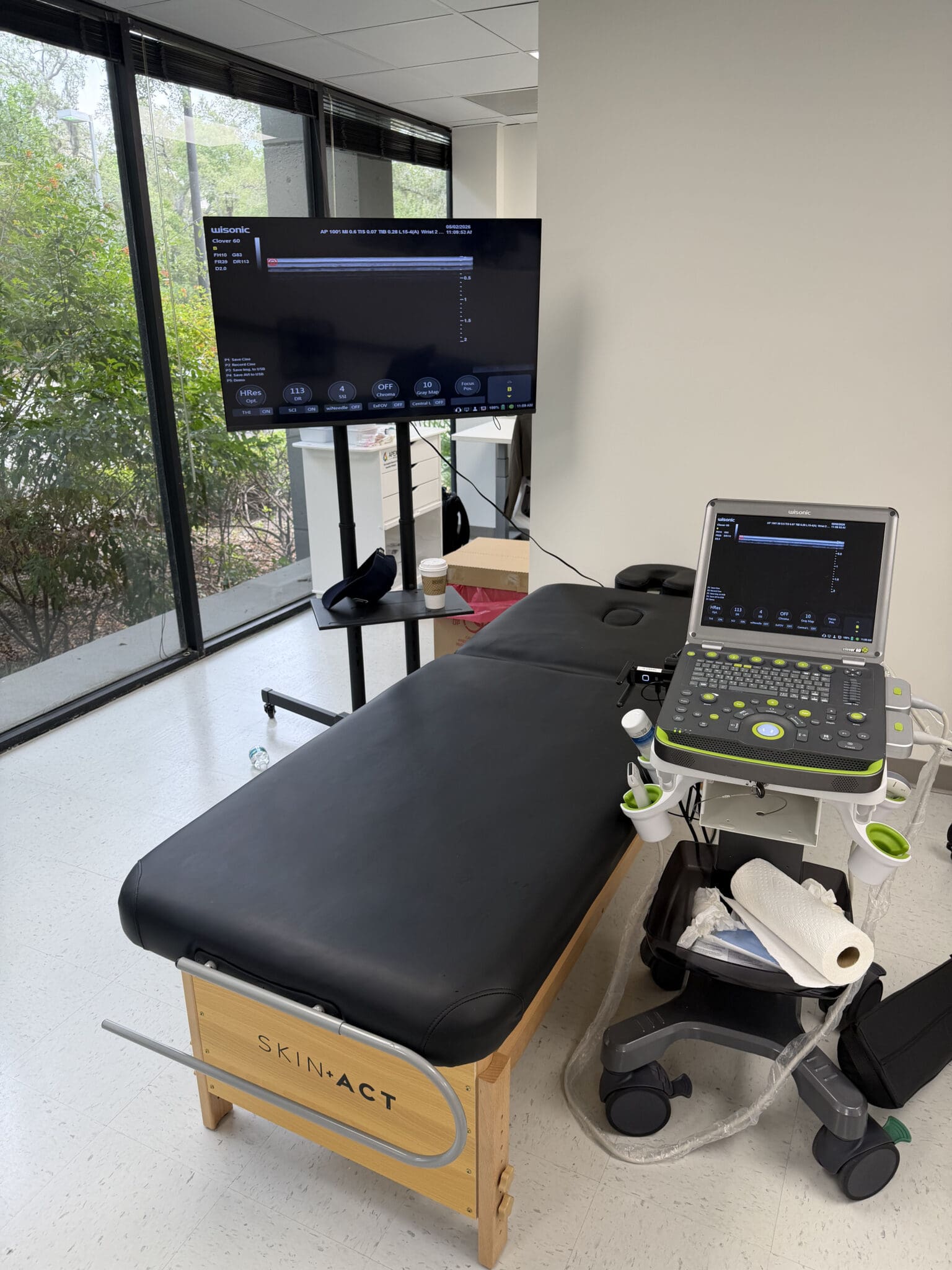
I prefer ultrasound guidance for intra-articular hip injections because it allows real-time visualization of critical anatomy and tissue planes. In this case, I use a 23-gauge needle for the admixture of PRP and plasma concentrate; if I were using plasma concentrate alone, I would opt for a 21-gauge due to viscosity.
Key steps I follow:
- I confirm the femoral head, acetabulum, and labrum in long axis to the neck/head.
- I identify the medial landmarking of the femoral artery pulsation to avoid the neurovascular bundle.
- I align the probe to be perpendicular to the femoral head surface to sharpen echogenic interfaces and maximize needle visualization.
Technical considerations and reasoning:
- Needle visualization is non-negotiable. I adjust the needle angle slightly steeper, if needed, so that the shaft and tip remain in plane on the superior portion of the image. Continuous visualization reduces the risk of extra-articular deposition and neurovascular contact (Finnoff et al., 2015).
- I purge all air from the line to prevent echogenic artifact that can obscure the tip.
- Pain feedback matters. If the patient reports disproportionate pain or I feel resistance, I reassess the tip location. Painful expansion suggests extra-articular or intratendinous placement. Intra-articular flow should feel smooth and appear as a clear anechoic spread within the joint recess.
- Flow profile: I watch for a “beautiful amount of fluid” to accumulate in the joint space, with minimal capsular tenting, indicating proper intra-articular dispersion.
I anesthetize the track beforehand and enter at a skin point that offers a straight shot to the anterior joint recess. With proper alignment, I slowly deliver the PRP mixture while continuously verifying the tip’s location.
What Patients Feel and What I Look For During Injection
Patient-reported soreness during needle advancement is common, but sudden sharp pain or pressure can indicate an off-target trajectory. During injection, I expect:
- Mild to moderate pressure that is tolerable
- Smooth syringe plunger depression without excessive force
- Clear sonographic spread within the joint space, sometimes with slight capsular bowing
If I perceive resistance, I pause. I may withdraw a millimeter, change the angle slightly, or re-scan to confirm the tip. The hip is unforgiving with volume and pressure, so finesse is key.
Post-injection, I reassess patient comfort, neurovascular status, and provide immediate guidance on movement, load limits, and icing strategies as indicated.
Integrative Chiropractic Care: Making the Biologics “Stick”
A biologic injection is one piece of a comprehensive care plan. As a chiropractor and nurse practitioner working integratively, my aim is to optimize the biomechanical environment so that the biological’s molecular benefits translate into durable function.
Core components I incorporate:
- Neuromuscular control of the hip complex
- Focus: gluteus medius/minimus for frontal plane control, deep external rotators for femoral head centration, and iliopsoas for sagittal balance.
- Why: Strong, well-timed abductors and rotators reduce anterior and anterosuperior femoral head translation during turnout, arabesque, and passé. This reduces shear at the labrum and capsular strain.
- Methods: Side-lying hip abduction with pelvic neutrality; isometric external rotation in varying degrees of flexion; controlled articular rotations to build end-range strength.
- Spinal-pelvic alignment and segmental joint care
- Focus: lumbopelvic rhythm, sacroiliac joint mechanics, and thoracic mobility.
- Why: Altered lumbopelvic rhythm can tilt the acetabulum and bias impingement earlier in the ROM arc. Restoring segmental motion can offload the hip.
- Chiropractic applications: Specific high-velocity, low-amplitude adjustments when indicated; mobilizations; and instrument-assisted soft tissue release to normalize tone and improve proprioception.
- Fascial continuity and kinetic-chain integration
- Focus: iliotibial band, tensor fasciae latae, rectus femoris, adductor longus, and oblique abdominal sling.
- Why: Tension asymmetries across fascial lines can pull the femoral head off-center at end range. Normalizing these lines enhances centration and reduces compensatory clicks.
- Techniques: Myofascial release, cupping for superficial glide, flossing bands for occlusion-based mobilization, and eccentric loading to reshape tendon-fascial stiffness.
- Foot and ankle mechanics
- Why: Excessive pronation or limited dorsiflexion propagates internal rotation moments and valgus drift up the chain, challenging hip stabilizers.
- Care: Foot intrinsic strengthening, ankle dorsiflexion mobilization, and forefoot control drills to dampen upstream stress.
- Motor control in dance-specific positions
- Why: Generic strengthening is insufficient if not contextualized to turnout, extensions, and transitions.
- Methods: Graded exposure to turnout with constraints, tempo control, and metronomic pacing to teach force distribution at end range.
- Load management and recovery dosing
- Why: Biologic signaling requires a window with reduced inflammatory load and microtrauma.
- Protocol: For the first 48–72 hours post-injection, keep volume low, avoid NSAIDs that can blunt the platelet cascade, and emphasize sleep quality, protein intake, and hydration. Gradual reintroduction of technical training follows a milestone-based plan.
These strategies are rooted in reducing nociceptive drive, optimizing femoral head centration, and ensuring that the labrum and capsule experience favorable mechanical signals during the PRP-mediated repair window (the Delphi consensus in hip preservation circles supports progressive motor control and activity modification).
The Science Behind PRP and Plasma Proteins in the Hip
- Cellular and molecular effects
- Platelets release alpha granules containing PDGF, TGF-β, VEGF, and other mediators that recruit reparative cells, stimulate matrix synthesis, and modulate inflammation (Dohan Ehrenfest et al., 2009).
- Plasma proteins such as fibrin and fibronectin form a provisional matrix, enhancing growth factor retention and cell adhesion.
- For capsular tissue, TGF-β can promote collagen synthesis, potentially improving tissue quality and tensioning, if loading is dosed correctly.
- Evidence in hip pathology
- PRP has shown promise in improving pain and function in intra-articular hip conditions, including early osteoarthritic changes and labral-related pain, particularly when delivered with image guidance (Chahla et al., 2021; Dwyer et al., 2021).
- Image guidance improves accuracy and outcomes by ensuring intra-articular deposition and reducing the risk of periarticular dispersion, which may dilute the effects (Sofka et al., 2005; Finnoff et al., 2015).
- Viscosity and gauge selection
- Plasma protein concentrates can be viscous; a larger bore (21-gauge) reduces shear forces and allows steady delivery. When mixed with PRP, viscosity decreases slightly, permitting the use of a 23-gauge needle. Lower shear preserves platelet integrity.
- Dose-volume rationale for the hip
- Smaller joints, or those with tight capsules, react to volume with pain and reflexive guarding. Concentrating biologics enables sufficient molecular signaling without causing pressure-related pain or leakage. The hip particularly benefits from high-potency, low-volume strategies.
Ultrasound Landmarks and Needle Strategy: Step-by-Step
To make this transparent for peers and reassuring for patients, here is how I approach the injection:
- Position and probe
- Supine, neutral hip. Anterior approach with a high-frequency linear transducer if body habitus permits; otherwise, a curvilinear probe.
- Identify femoral head-neck junction and acetabular rim; confirm labrum as a hyperechoic triangular structure.
- Safety scan
- Sweep medially to visualize femoral artery pulsation and vein; choose a skin entry lateral enough to avoid the neurovascular bundle.
- Needle plan
- In-plane approach, advancing toward the anterior joint recess. Slightly steeper angle if visualization fades.
- Maintain the needle within the imaging plane; adjust probe tilt to keep the tip bright and crisp.
- Confirmation
- Test with a minuscule saline bubble if needed to confirm spread. Then deliver the PRP-protein mixture slowly, watching the anechoic collection expand intra-articularly.
- Troubleshooting
- If resistance or patient pain spikes, reassess tip position. Withdraw slightly, alter trajectory, or pause and rescan.
- Aftercare
- Gentle ROM later the same day, avoiding extremes. Ice for comfort as tolerated. Educate on expected soreness and red flags.
This methodical approach reduces variability and enhances outcomes by fostering precise placement and minimizing unnecessary tissue irritation.
Integrative Rehab Timeline After Hip PRP: What I Recommend
I use a phase-based timeline, individualized to tissue response. For a procedure performed on 2026-03-07:
- Phase 0: Days 0–3 (2026-03-07 to 2026-03-10)
- Priorities: Protect, reduce excessive load, and allow early anabolic signaling.
- Actions: Relative rest, gentle pain-free ROM, diaphragmatic breathing, pelvic tilts, isometric glute sets, short-lever abductor isometrics. Avoid NSAIDs; consider acetaminophen if needed.
- Phase 1: Days 4–14 (2026-03-11 to 2026-03-21)
- Priorities: Re-establish neuromuscular control, improve capsular mechanics without overstretch.
- Actions: Progress isometrics to short-range concentrics; begin controlled articular rotations; integrate foot-ankle stability drills; soft tissue work for TFL/rectus femoris if hypertonic; chiropractic mobilizations for lumbopelvic segments as indicated.
- Phase 2: Weeks 3–6 (2026-03-22 to 2026-04-18)
- Priorities: Strength and endurance in functional planes; graded exposure to turnout and extensions.
- Actions: Hip abduction/ER in closed chain, lateral step-downs with pelvis control, anti-rotation core drills, tempo-based eccentric loading, introduction of dance-specific patterns at submax intensity.
- Phase 3: Weeks 7–10 (2026-04-19 to 2026-05-24)
- Priorities: Power, control at end range, and return to choreography complexity.
- Actions: Plyometric progressions, complex transitions with fatigue-resistance, sport-specific constraints to maintain centration at maximal ROM. Continue targeted manual therapy as needed.
- Phase 4: Beyond Week 10 (from 2026-05-25 forward)
- Priorities: Sustain capacity; periodic deloads; maintenance care.
- Actions: Load cycling, recovery monitoring, occasional booster neuromuscular sessions, and re-evaluation of hip mechanics under performance stress.
This sequencing respects biologic healing windows while reconditioning the neuromuscular system to support long-term joint health.
Clinical Observations From Practice: Patterns I See and Correct
Across my clinical work, including cases shared on my professional outlets, several patterns recur in dancers with hypermobility and hip pain:
- Overreliance on anterior structures
- TFL dominance with underactive posterior gluteal chain. Outcome: Anterior capsular stress and labral compression.
- Correction: Posterior chain activation, cueing pelvis-neutral abduction, and stride mechanics emphasizing heel-toe progressions to encourage hip extension without lumbar substitution.
- Thoracolumbar extension bias
- Dancers often compensate for hip extension limits with lumbar extension.
- Correction: Thoracic mobility plus core anti-extension drills, allowing true hip extension without shear.
- Foot control deficits
- Midfoot collapse increases internal rotation moments.
- Correction: Short-foot training, tripod stance drills, and resisted inversion-eversion control to refine ground-up stability.
- Fatigue-induced form drift
- Technique erodes late in sessions; hip centration fails first.
- Correction: Use of metronomic pacing and time-capped sets with quality thresholds; build load tolerance gradually.
When biologics are integrated into this framework, I observe greater durability of symptom relief and functional restoration. The injection calms the storm; the integrative program rebuilds the harbor.
You can see more of my clinical perspectives and case insights here:
- Clinical observations and resources: https://pushasrx.com/
- Professional background and updates: https://www.linkedin.com/in/dralexjimenez/
Risks, Expectations, and Informed Decision-Making
Any injection carries risks: infection, bleeding, neurovascular injury, transient pain flares, or failure to improve. With ultrasound guidance, risk is reduced by visualizing critical structures. For PRP, patients may experience a temporary increase in soreness as inflammatory signaling initiates. I advise on red flags, set realistic expectations, and align the biologic intervention with a comprehensive plan that addresses movement quality and tissue capacity.
The anticipated benefits include improved pain, better tolerance of end-range positions, and reduced clicking if driven by microinstability rather than a macro labral tear. If a significant labral tear is later identified or conservative-integrative care is insufficient, I coordinate with hip preservation surgeons for further evaluation, ensuring continuity of care.
Putting It All Together: A Patient-Centered, Performance-Ready Hip
- Diagnose accurately with imaging and functional testing.
- Use precise ultrasound-guided biologic delivery to target intra-articular pathology while respecting hip volume sensitivity.
- Implement integrative chiropractic care to normalize joint mechanics and motor control.
- Progress loading with intention, matching the biologic healing curve.
- Monitor outcomes and adapt the plan with the patient’s goals front and center.
For this dancer, the combination of high-concentration PRP plus plasma protein concentrate—delivered safely and accurately—provides a biologic platform for recovery. The integrative plan ensures that as tissues heal, the system learns to move better, stronger, and more sustainably at the edge of performance.
References
- Chahla, J., et al. (2021). Platelet-rich plasma for joint preservation in orthopedics: evidence and recommendations. Arthroscopy, 37(7), 2262–2275.
- Dohan Ehrenfest, D. M., et al. (2009). Classification of platelet concentrates: from pure platelet-rich plasma (P-PRP) to leucocyte- and platelet-rich fibrin (L-PRF). Trends in Biotechnology, 27(3), 158–167.
- Dwyer, T., et al. (2021). The role of biologics in hip preservation surgery and nonoperative care. Clinics in Sports Medicine, 40(2), 331–350.
- Everts, P., et al. (2020). Autologous platelet-rich plasma and platelet-rich fibrin in orthopedics and sports medicine. Muscles, Ligaments and Tendons Journal, 10(3), 322–336.
- Finnoff, J. T., et al. (2015). Accuracy of ultrasound-guided versus unguided intra-articular injections. PM&R, 7(4), 378–384.
- Kalisvaart, M. M., & Safran, M. R. (2015). Hip instability treated with arthroscopic capsular plication. Knee Surgery, Sports Traumatology, Arthroscopy, 24(12), 3746–3751.
- LaFrance, R. M., et al. (2021). Outcomes of nonoperative treatment for hip labral tears and femoroacetabular impingement. Sports Health, 13(3), 191–200.
- Nepple, J. J., et al. (2015). The hip fluid seal—Part I: the effect of an acetabular labral tear, repair, resection, and reconstruction on hip fluid pressurization. Knee Surgery, Sports Traumatology, Arthroscopy, 23(10), 3067–3073.
- Sofka, C. M., et al. (2005). Ultrasound-guided interventions of the hip and pelvis. AJR American Journal of Roentgenology, 184(2), 547–551.
- Fitzpatrick, J., et al. (2017). The effectiveness of platelet-rich plasma in the treatment of tendinopathy: a meta-analysis. American Journal of Sports Medicine, 45(1), 226–233.
Post Disclaimer *
Professional Scope of Practice *
The information herein on "Ultrasound PRP for Hip Impingement Recovery Process" is not intended to replace a one-on-one relationship with a qualified health care professional or licensed physician and is not medical advice. We encourage you to make healthcare decisions based on your research and partnership with a qualified healthcare professional.
Blog Information & Scope Discussions
Welcome to El Paso's Premier Fitness, Injury Care Clinic & Wellness Blog, where Dr. Alex Jimenez, DC, FNP-C, a Multi-State board-certified Family Practice Nurse Practitioner (FNP-BC) and Chiropractor (DC), presents insights on how our multidisciplinary team is dedicated to holistic healing and personalized care. Our practice aligns with evidence-based treatment protocols inspired by integrative medicine principles, similar to those found on this site and our family practice-based chiromed.com site, focusing on restoring health naturally for patients of all ages.
Our areas of multidisciplinary practice include Wellness & Nutrition, Chronic Pain, Personal Injury, Auto Accident Care, Work Injuries, Back Injury, Low Back Pain, Neck Pain, Migraine Headaches, Sports Injuries, Severe Sciatica, Scoliosis, Complex Herniated Discs, Fibromyalgia, Chronic Pain, Complex Injuries, Stress Management, Functional Medicine Treatments, and in-scope care protocols.
Our information scope is multidisciplinary, focusing on musculoskeletal and physical medicine, wellness, contributing etiological viscerosomatic disturbances within clinical presentations, associated somato-visceral reflex clinical dynamics, subluxation complexes, sensitive health issues, and functional medicine articles, topics, and discussions.
We provide and present clinical collaboration with specialists from various disciplines. Each specialist is governed by their professional scope of practice and their jurisdiction of licensure. We use functional health & wellness protocols to treat and support care for musculoskeletal injuries or disorders.
Our videos, posts, topics, and insights address clinical matters and issues that are directly or indirectly related to our clinical scope of practice.
Our office has made a reasonable effort to provide supportive citations and has identified relevant research studies that support our posts. We provide copies of supporting research studies upon request to regulatory boards and the public.
We understand that we cover matters that require an additional explanation of how they may assist in a particular care plan or treatment protocol; therefore, to discuss the subject matter above further, please feel free to ask Dr. Alex Jimenez, DC, APRN, FNP-BC, or contact us at 915-850-0900.
We are here to help you and your family.
Blessings
Dr. Alex Jimenez DC, MSACP, APRN, FNP-BC*, CCST, IFMCP, CFMP, ATN
email: [email protected]
Multidisciplinary Licensing & Board Certifications:
Licensed as a Doctor of Chiropractic (DC) in Texas & New Mexico*
Texas DC License #: TX5807, Verified: TX5807
New Mexico DC License #: NM-DC2182, Verified: NM-DC2182
Multi-State Advanced Practice Registered Nurse (APRN*) in Texas & Multi-States
Multistate Compact APRN License by Endorsement (42 States)
Texas APRN License #: 1191402, Verified: 1191402 *
Florida APRN License #: 11043890, Verified: APRN11043890 *
Verify Link: Nursys License Verifier
* Prescriptive Authority Authorized
ANCC FNP-BC: Board Certified Nurse Practitioner*
Compact Status: Multi-State License: Authorized to Practice in 40 States*
Graduate with Honors: ICHS: MSN-FNP (Family Nurse Practitioner Program)
Degree Granted. Master's in Family Practice MSN Diploma (Cum Laude)
Dr. Alex Jimenez, DC, APRN, FNP-BC*, CFMP, IFMCP, ATN, CCST
(Board Certified: Family Practice Nurse Practitioner—Multistate)*
(Licensed Nurse Practitioner & Chiropractor - Multistate)*
Clinical Director
Digital Business Card
Dr. Maria Cardenas, MD
(Board Certified: Internal Medicine)
(Licensed Medical Doctor)
Medical Director, Clinical Director & Collaborative Physician
NPI # 1164426749
MD License #: J2933
Licenses and Board Certifications:
MD: Medical Doctor
DC: Doctor of Chiropractic
APRNP: Advanced Practice Registered Nurse
FNP-BC: Family Practice Specialization (Multi-State Board Certified)
RN: Registered Nurse (Multi-State Compact License)
CFMP: Certified Functional Medicine Provider
MSN-FNP: Master of Science in Family Practice Medicine
MSACP: Master of Science in Advanced Clinical Practice
IFMCP: Institute of Functional Medicine
CCST: Certified Chiropractic Spinal Trauma
ATN: Advanced Translational Neutrogenomics
Memberships & Associations:
TCA: Texas Chiropractic Association: Member ID: 104311
AANP: American Association of Nurse Practitioners: Member ID: 2198960
ANA: American Nurse Association: Member ID: 06458222 (District TX01)
TNA: Texas Nurse Association: Member ID: 06458222
NPI: 1205907805
| Primary Taxonomy | Selected Taxonomy | State | License Number |
|---|---|---|---|
| No | 111N00000X - Chiropractor | NM | DC2182 |
| Yes | 111N00000X - Chiropractor | TX | DC5807 |
| Yes | 363LF0000X - Nurse Practitioner - Family | TX | 1191402 |
| Yes | 363LF0000X - Nurse Practitioner - Family | FL | 11043890 |
| Yes | 363LF0000X - Nurse Practitioner - Family | CO | C-APN.0105610-C-NP |
| Yes | 363LF0000X - Nurse Practitioner - Family | NY | N25929 |
Dr. Alex Jimenez, DC, APRN, FNP-BC*, CFMP, IFMCP, ATN, CCST
(Board Certified: Family Practice Nurse Practitioner—Multistate)*
(Licensed Nurse Practitioner & Chiropractor - Multistate)*
Clinical Director
Digital Business Card
Dr. Maria Cardenas, MD
(Board Certified: Internal Medicine)*
(Licensed Medical Doctor)*
Medical Director, Clinical Director & Collaborative Physician
NPI # 1164426749
MD License #: J2933